Fluorescence in the second near-infrared (NIR-II) region with wavelengths from 1000 to 1400 nm has exhibited great promising in in vivo epifluorescence imaging, in comparison to that in the first NIR window (NIR-I) with wavelengths from 650 to 950 nm, due to the much reduced photon absorption and scattering by tissues, which offers deeper tissue penetration and higher spatial and temporal resolution. Up to date, the most promising NIR-II emissive nanoprobes are limited to single-walled carbon nanotubes (SWNTs) and Ag2S quantum dots (QDs). Studies have indicated that the NIR-II emission centered at 1300 nm is the optimal wavelength for in vivo imaging with the deepest tissue penetration due to the lowest photon absorption and tissue scattering.Although CdHgTe, PbS, and PbSe QDs have been previously reported, however, the intrinsic toxicity of the heavy metal elements deters their potential practice in in vivo imaging. Therefore, it is urgent to develop a new type of bright and biocompatible NIR-II nanoprobe with an emission wavelength centered at 1300 nm for in vivo deep tissue/organ imaging.
Prof. WANG Qiangbin’s group from Suzhou Institute of Nano-Tech & Nano-Bionics, CAS, has prepared NIR-II nanoprobe, Ag2S QDs, (J. Am. Chem. Soc. 2010, 132, 1470–1471), with emission centered at 1200 nm, which have been successfully employed in cellular imaging and xenograft tumors imaging with a high signal-to-noise ratio (ACS Nano 2012, 6, 3695-3702, Angew. Chem. Int. Ed. 2012, 51, 9818-9821).
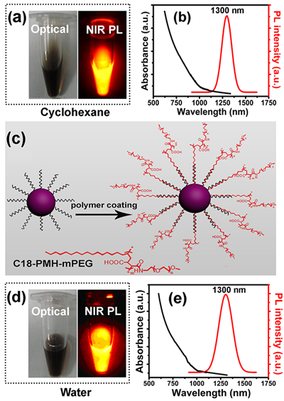
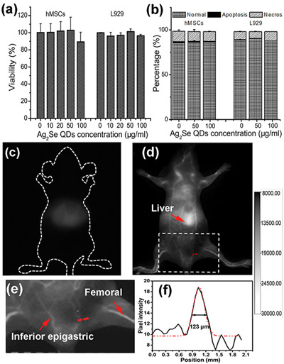
Recently, they develop a new tape of NIR-II highly photoluminescent Ag2Se QDs with emission at 1300 nm. Functionalizing the surface with an amphiphilic C18-PMH-PEG polymer, the obtained C18-PMH-PEG-Ag2Se QDs exhibit bright NIR-II fluorescence, good water solubility, high colloidal stability and photostability, as well as decent biocompatibility. The in vivo experiment by administration of C18-PMH-PEG-Ag2Se QDs by tail intravenous injection achieves high spatial resolution imaging of organs buried deep in the body and vascular structures down to ~100 μm. This new NIR-II fluorescent nanoprobe with small sizes, ideal optical properties, and decent biocompatibility opens up exciting opportunities for future biomedical applications. This work has been recently published in Chem. Mater. 2013, 25, 2503-2509.
This work was supported by “Hundred Talents” program of CAS, Strategic Priority Research Program of CAS, NSFC, and MOST.

Figure 1. Optical properties of Ag2Se QDs before (a, b) and after (d, e) C18-PMH-PEG coating.(Image by SINANO)

Figure 2.Cytotoxicity assay (a, b) and in vivo imaging (c-f) of C18-PMH-PEG-Ag2Se QDs.(Image by SINANO)
downloadFile
