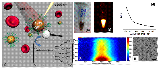
With the development of biomedical imaging, near infrared fluorescence imaging has drawn intensive attention in the field of biomedicine. Among these techniques, fluorescent imaging in the second near-infrared window (NIR-II, 1.0-1.4 μm) is appealing due to minimal autofluorescence and negligible tissue scattering, affording maximal penetration depth for deep tissue imaging with high feature fidelity. Quantum dots (QDs), as new fluorescent nanoprobes, have been widely used in biological labeling, sensing, and imaging due to unique advantages such as high brightness, excellent photostability, spectral tunability. Development of biocompatible NIR-II QDs with high quantum yield for in vivo imaging remains a challenge.
Prof. WANG Qiangbin’s group from Suzhou Institute of Nano-Tech & Nano-Bionics, CAS, has prepared NIR-II Ag2S QDs with higher quantum yield, better biocompatibility, and controllable sizes based on their previous work, “Near-Infrared Photoluminescent Ag2S Quantum Dots from a Single Source Precursor” (J. Am. Chem. Soc. 2010, 132, 1470–1471). They also performed cellular imaging studies using Ag2S QDs and evaluation of Ag2S QD cytotoxicity, in collaboration with DAI HongJie’s team at Stanford University. They demonstrated that cell-specific lableling and imaging could be achieved using Ag2S QDs conjugated with targeting ligands and that Ag2S QDs showed negligible cytotoxicity (ACS Nano 2012, 6, 3695–3702).
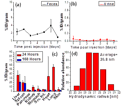
Then they continued to develop Ag2S QDs for in vivo imaging in animals. It has been found that tumor has a high uptake of Ag2S QDs (Figure 2) because of the enhanced permeability and retention (EPR) effect of tumor vasculature, which provides an important technical foundation for the early diagnosis of tumor and surgery visualization. They also studied the short-term retention and excretion after a single-dose injection of the Ag2S QDs into tumor-free BALB/c mice (n=4). Feces and urine are collected on a daily basis for quantitative measurement of Ag2S QDs based on ICP-MS. They found that Ag2S QDs were nearly cleared from all organs aside from the liver and the spleen after 7 days (Figure 3).This work has been recently published in Angewandte Chemie International Edition.
This work was supported by “Hundred Talents” program of CAS, Strategic Priority Research Program of CAS, NSFC, and MOST.