Spinal cord injury (SCI) repair has remained one of the most challenging clinical problems. Over recent years, neural progenitor cells (NPCs) are considered to be promising in cell replacement strategies. The main challenge for neural progenitor cell (NPC)-mediated repair of spinal cord injury (SCI) is lack of favorable environment to direct NPCs differentiation towards neurons rather than glial cells. The myelin associated inhibitors at the lesion site have been demonstrated to promote NPC differentiation into glial lineage.
Recently, Dr. DAI Jianwu’s research group at Suzhou Institute of Nano-Tech & Nano-Bionics, Chinese Academy of Sciences(SINANO), reported an EGFR antibody functionalized collagen scaffold to promote neuronal differentiation of neural progenitor cells
In this work, cetuximab, an epidermal growth factor receptor (EGFR) neutralizing antibody, was used to inhibit the downstream signaling activated by myelin associated inhibitors and to functionalize collagen scaffold for NPC implantation. It showed that collagen-cetuximab 1 µg scaffolds enhanced neuronal differentiation and inhibited astrocytic differentiation of NPCs exposed to myelin proteins significantly in vitro. To test the therapeutic effect in vivo, NPCs expressing green fluorescent protein (GFP)-embedded scaffolds have been implanted into the 4 mm-long hemisection lesion of rats. It was found that the collagen-cetuximab 5 µg scaffolds induced neuronal differentiation and decreased astrocytic differentiation of NPCs, enhanced axon regeneration, and promoted functional recovery markedly. This work was published in Biomaterial (2013, 34, 5107).
This work was supported by grants from the Ministry of Science and Technology of China and National Science Foundation of China.
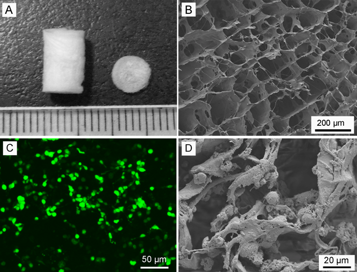
Fig. 1. (A) Picture displayed the structure of the collagen scaffold, (B) SEM image, (C) Confocal image of FDA staining and (D) SEM image of NPCs cultured on collagen scaffold for 7 days.(Image by SINANO)
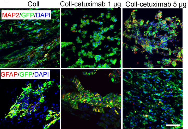
Fig. 2. Differentiation of NPCs in hemi-transection site at12 weeks post-SCI in rat.(Image by SINANO)