Intravital fluorescence-based optical imaging has inherent advantages owing to its high sensitivity, fast feedback, multiplexing, and absence of ionizing radiation. Fluorescence imaging in the second near-infrared region (NIR-II, 1000–1350 nm) is more desirable than visible (450–700 nm) and traditional NIR-I imaging (700–950 nm) owing to greatly reduced photon absorption and scattering by tissues, as well as negligible tissue autofluorescence, which promises high-fidelity imaging of deeper tissues and organs. Recently, Prof. Wang Qiangbin’s group from Suzhou Institute of Nano-Tech & Nano-Bionics, CAS(SINANO), has reported that Ag2S quantum dots (QDs), containing no toxic ions, exhibiting long circulation time and high stability, act as a new kind of fluorescent probes in the second near-infrared window (NIR-II, 1000-1350 nm) which enable in vivo monitoring of lymphatic drainage and vascular networks with deep tissue penetration and high spatial and temporal resolution. In addition, NIR-II fluorescence imaging with Ag2S QDs provide ultrahigh spatial resolution (~40 mm) that permits to track angiogenesis mediated by a tiny tumor (2-3 mm in diameter) in vivo. Their results indicate that Ag2S QDs are promising NIR-II fluorescent nanoprobes that could be useful in surgical treatments such as sentinel lymph node (SLN) dissection as well in assessment of blood supply in tissues and organs and screening of anti-angiogenic drugs. This work has been published in Biomaterials 2014, 35, 393-400. This work was supported by “Hundred Talents” program of CAS, Strategic Priority Research Program of CAS, NSFC, and MOST. Video 1: NIR-II video-rate imaging of blood flow in the nude mouse after injected with PEGylated Ag2S QDs (200 μL, 1mg/mL).(Video by SINANO) 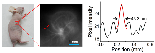
Figure 1. In vivo imaging of tumor-induced angiogenesis with Ag2S QDs (1200 nm, NIR-II).(Image by SINANO) 
Figure 2. In vivo imaging of the lymphatic system with ICG (835 nm, NIR-I) and Ag2S QDs (1200 nm, NIR-II).(Image by SINANO) |