Mesenchymal stem cells (MSCs) have shown great potential for cutaneous wound regeneration in clinical practice. However, the in vivo homing behavior of intravenously transplanted MSCs to the wounds is still poorly understood. Thus, the development of high-resolution imaging techniques to monitor the temporal and spatial homing of transplanted stem cells to target tissues in living animal model is urgently needed for a better understanding of both the therapeutic mechanisms and the safety issues of stem cells before their uses in clinical trials.
Recently, a team led by Prof. Wang Qiangbin from Suzhou Institute of Nano-Tech & Nano-Bionics, Chinese Academy of Sciences (SINANO), developed an Ag2S QDs-based NIR-II imaging technique for in vivo visualizing the dynamic homing behavior of transplanted human mesenchymal stem cells (hMSCs) to a cutaneous wound in mice. This work was recently published in Biomaterials 2015, 53, 265-273.
Benefiting from the desirable spatial and temporal resolution of Ag2S QDs-based NIR-II imaging, for the first time, the migration of hMSCs to the wound was dynamically visualized in vivo. For the wound treated by the collagen scaffold loaded with stromal cell derived factor-1α (SDF-1α), more hMSCs were recruited at the wound within a much shorter time and were homogenously distributed across the whole wound area, which enhances the re-epithelialization, the neovascularization, and accelerates the wound healing. Their findings offer direct evidence of the important roles of hMSCs and SDF-1a involved in the cutaneous regeneration, and help to disclose the underlying mechanism of stem cell-based regeneration study.
This work was supported by the Chinese Academy of Sciences “Strategic Priority Research Program”, the Ministry of Science and Technology of China, the National Natural Science Foundation of China and the National Natural Science Foundation of Jiangsu Province.
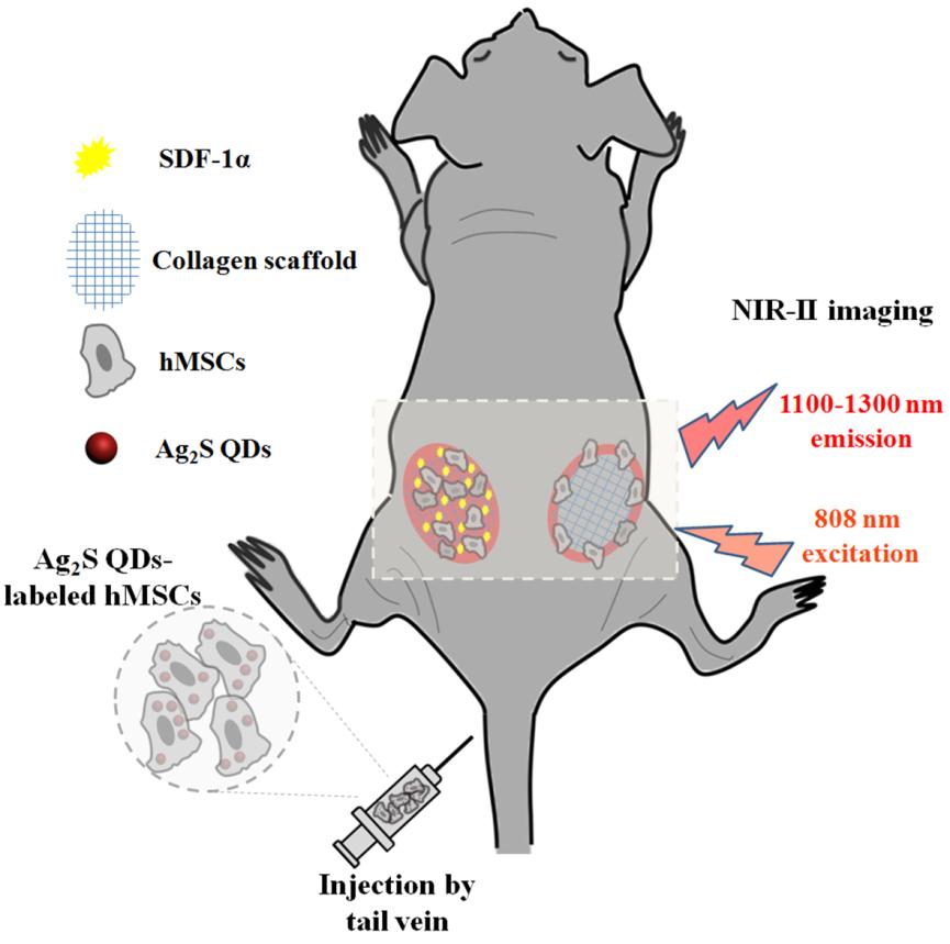
Figure 1. Schematic of Ag2S QDs-based NIR-II imaging for in vivo tracking of MSCs tropism for cutaneous regeneration.(Image by Prof. Wang Qiangbin's group)
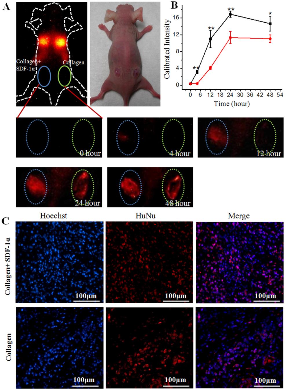
Figure 2. In vivo visualization of the migration of hMSCs. (A) Photoluminescence images of the migration of hMSCs. Left, wound filled with SDF-1α-loaded collagen; right, wound filled with blank collagen fragments. (B) Fluorescence intensity of Ag2S QDs-labeled hMSCs accumulated in the wound. (C) Immunofluorescence staining with Hoechst (blue) for all cells and HuNu (red) for hMSCs.(Image by Prof. Wang Qiangbin's group)