Stem cell-based regenerative medicine holds great promising in clinic practices. However, the stem cells after transplantation experience a very different microenvironment from in vitro and their viability in vivo is critical to the therapeutic effect. Till now, the fate of transplanted stem cells, including the distribution, viability and the cell clearance by the immune system, has not been fully understood yet. Thus, a non-invasive in vivo imaging technique that can monitor the fate of transplanted stem cells is urgently needed for deeper understanding the role of stem cell played in regenerative medicine and therefore speeding up the clinical translation of stem cell-based therapeutics.
Recently, Prof. WANG Qiangbin’s group from Suzhou Institute of Nano-Tech and Nano-Bionics (SINANO), Chinese Academy of Sciences (CAS), developed a dual-labeling strategy to in situ visualize the fate of transplanted stem cells in vivo by combining the exogeneous near-infrared fluorescence imaging (NIRFI) of Ag2S quantum dots (emitting at 1200 nm) and endogenous bioluminescence imaging (BLI) of red-emitting firefly luciferase (RfLuc, emitting at 613 nm), due to the deep tissue penetration and high spatiotemporal resolutin of NIRFI and the characteristic nature of living stem cells to express RfLuc (Figure). For the first time, researchers have successfully traced the dynamic translocation of mMSCs in vivo, differentiated the living cells and dead cell components in situ, and further clarified the regenerative mechanism of living mMSCs involved in the healing of the acute liver failure by using the RfLuc/Ag2S QDs dual-label method. The distinct features of the dual-imaging method encourge a broad range of further applications, such as in vivo stem cell intervention, imaging-guided cell therapeutics, and so on. These findings can inspire future stem cell research and this novel noninvasive imaging method is promising in future biomedicine studies and will provide us more fundamental understanding on the in vivo biological activities.
This study was recently published in Small under the title “Revealing the Fate of Transplanted Stem Cells In Vivo with a Novel Optical Imaging Strategy”.
This work was supported by the National Key Research and Development Program, the National Natural Science Foundation of China, and the Natural Science Foundation of Jiangsu Province.
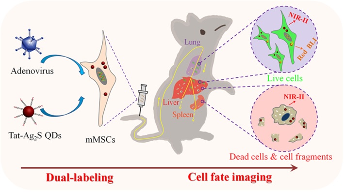
Figure: Schematic illustration of the dual-labeling strategy for in vivo tracking the fate of transplanted stem cells by combining the exogenous Tat-Ag2S QDs and endogenous RfLuc (Image by WANG Qiangbin and CHEN Guangcun)
(Nanjing Branch of Chinese Academy of Sciences)